


The article ‘Eye Anatomy: Parts of the Human Eye detailed explained’ provides a comprehensive overview of the intricate structure and functions of the human eye. It explores key components such as the cornea, lens, retina, and optic nerve, elucidating their roles in vision and visual perception. Additionally, it delves into topics like the mechanics of light refraction, the significance of the visual processing center in the brain, and various eye conditions and surgical interventions. Furthermore, it examines the muscles, movements, and auxiliary structures of the eye, emphasizing their contribution to eye health. Finally, it touches upon aspects of vision and perception, including color, depth, and integration.
Overview of Eye Anatomy
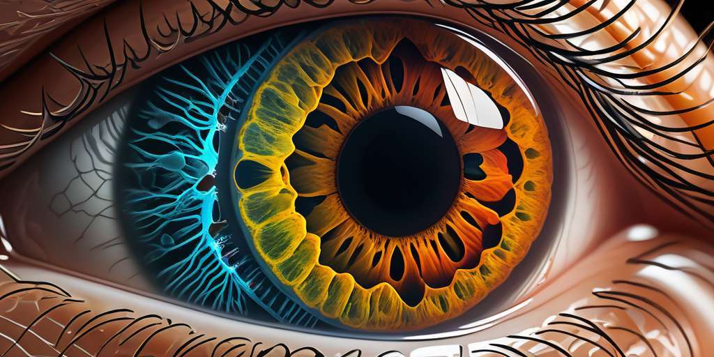
The human eye is a remarkable organ responsible for our sense of vision. Understanding its anatomy is essential for comprehending how it functions and enables us to see the world around us.
The eye consists of several interconnected parts, each with a specific role in the visual process. Starting from the front, we have the cornea, which acts as a protective outer layer and helps focus incoming light. Behind the cornea lies the lens, which further refracts light to create a clear image on the retina.
The retina, located at the back of the eye, is a thin layer of tissue containing specialized cells called photoreceptors. These cells convert light into electrical signals that are transmitted to the brain via the optic nerve.
In addition to these main components, the eye also includes structures like the conjunctiva and eyelids, which protect the eye and lubricate its surface, and various muscles that control eye movements.
Understanding the anatomy of the eye provides a foundation for exploring topics such as light refraction, vision disorders, and surgical interventions. It sheds light on how we perceive colors, depth, and integrate visual information in our brain. By delving into the intricate details of eye anatomy, we gain a deeper appreciation for the wonders of vision and the complexity of the human eye.
The Structure and Function of the Cornea
The cornea, located at the front of the eye, plays a crucial role in our vision. It is a transparent, dome-shaped tissue that acts as a protective outer layer for the eye. The cornea refracts light, bending it as it enters the eye, and helps to focus it onto the retina at the back. Its smooth surface and curvature are essential for maintaining clear and sharp vision.
The cornea consists of several different layers. The outermost layer, called the epithelium, protects the cornea from external elements and controls the amount of moisture on its surface. Beneath the epithelium is the stroma, which forms the majority of the cornea and provides strength and support. Finally, the innermost layer, known as the endothelium, helps maintain the clarity of the cornea by regulating fluid balance.
With its remarkable transparency, the cornea allows light to pass through easily. It also lacks blood vessels, relying on the tears and aqueous humor to provide it with nutrients and oxygen. The cornea’s unique structure and function contribute to its ability to focus light accurately onto the retina, enabling us to perceive the world around us.
Key points:
- The cornea refracts light and helps focus it onto the retina.
- It consists of the epithelium, stroma, and endothelium layers.
- The cornea is transparent and lacks blood vessels.
- Tears and aqueous humor provide nutrients and oxygen to the cornea.
Understanding the Role of the Lens in Vision
The lens is a crucial component of the human eye that plays a vital role in vision. It is a transparent, flexible structure located behind the iris and is responsible for focusing light onto the retina. The lens works together with the cornea to bend and refract incoming light, ensuring that it converges precisely on the retina.
Through a process called accommodation, the lens changes its shape to adjust the focus based on the distance of the object being observed. This versatile adjustment allows for clear vision at various distances, whether it’s nearby objects or objects in the distance.
The lens also plays a significant role in helping to control the amount of light entering the eye. It helps to regulate the size of the pupil, which determines the amount of light that reaches the retina. This mechanism ensures that the retina receives an optimal amount of light for clear and comfortable vision.
The Importance of the Retina in Visual Perception
The retina plays a crucial role in visual perception, acting as the light-sensitive layer that captures images and transmits them to the brain for interpretation. This thin layer of tissue lines the back of the eye and contains specialized cells called photoreceptors.
Within the retina, there are two types of photoreceptors: rods and cones. Rods are responsible for detecting light and shades of gray, enabling us to see in low light conditions. On the other hand, cones are responsible for perceiving colors and details in bright light.
When light enters the eye, it passes through the other layers of the eye, such as the cornea and lens, before reaching the retina. Once the light reaches the retina, it interacts with the photoreceptor cells, triggering a cascade of chemical reactions that convert the light into electrical signals.
These electrical signals are then transmitted to the optic nerve, which sends them to the visual processing center in the brain for interpretation. Through complex neural pathways, the brain processes these signals and constructs the visual images we perceive, allowing us to recognize shapes, colors, and objects in our environment.
In summary, the retina acts as the crucial interface between light entering the eye and the brain’s interpretation of visual stimuli. Without a healthy and properly functioning retina, our ability to perceive the world around us would be significantly impaired.
Exploring Vision Center: How the Eye Transmits Signals to the Brain
Within the complex network of our visual system, the eye plays a crucial role in transmitting signals to the brain for interpretation. It’s fascinating how this intricate process occurs seamlessly, allowing us to perceive the world around us.
The journey begins with the retina, a thin layer of tissue at the back of the eye that contains specialized cells called photoreceptors. These cells, known as rods and cones, convert light into electrical signals.
Once these signals are generated, they travel through the optic nerve, a bundle of more than a million nerve fibers. The optic nerve carries the visual information to the brain’s visual cortex, located in the occipital lobe. Here, the brain processes and interprets the signals to form a visual perception.
It’s important to note that the information transmitted to the brain is not a replica of the physical world but rather a representation constructed by the brain itself. This processing involves the integration of visual cues, such as color, motion, and depth, to create a complete image.
Understanding how the eye transmits signals to the brain is essential in comprehending the intricacies of human vision. This process allows us to experience the richness and beauty of the visual world that surrounds us.
The Significance of Light in the Visual Process
Light plays a fundamental role in the visual process, allowing us to perceive and understand the world around us. The human eye is exquisitely adapted to capture and interpret light, converting it into electrical signals that can be processed by the brain.
When light enters the eye through the cornea, it undergoes refraction, which is the bending of light rays to focus them onto the retina. The lens further adjusts the focus to ensure a clear image formation on the retina.
Once the light reaches the retina, it encounters specialized cells called photoreceptors, namely rods and cones. The rods are responsible for vision in low-light conditions, while the cones enable us to perceive color and fine details.
When light stimulates the photoreceptor cells, they undergo a chemical reaction that generates electrical signals. These signals are then transmitted through the optic nerve to the visual processing center in the brain, where they are interpreted as visual images.
Understanding the significance of light in the visual process is crucial for comprehending the mechanisms underlying vision and various eye conditions. By studying the interactions between light, the eye’s anatomy, and the brain, researchers and healthcare professionals can develop a better understanding of how to diagnose, treat, and prevent visual impairments.
Common Eye Conditions and Anatomy Related to Vision Problems
The human eye is vulnerable to various conditions and anatomical factors that can affect vision. Understanding these common eye conditions and their related anatomy is crucial for maintaining eye health and seeking appropriate treatment.
- Myopia (Nearsightedness): This condition occurs when the eyeball is too elongated or the cornea is too curved, causing light to focus in front of the retina instead of directly on it.
- Hyperopia (Farsightedness): In contrast to myopia, hyperopia occurs when the eyeball is too short or the cornea is too flat, causing light to focus behind the retina.
- Astigmatism: This common condition results from an irregularly shaped cornea, which leads to distorted vision at all distances.
- Presbyopia: As people age, the lens of the eye loses flexibility, making it difficult to focus on near objects. This condition is known as presbyopia.
In addition to these refractive errors, several eye diseases can also impact vision:
- Cataracts: Cataracts occur when the lens becomes clouded, resulting in blurred or hazy vision.
- Glaucoma: Glaucoma is a group of eye conditions that cause damage to the optic nerve, often due to increased pressure within the eye.
- Macular degeneration: This condition involves the deterioration of the central portion of the retina, known as the macula, leading to a loss of central vision.
Understanding these common eye conditions, along with the underlying anatomy and mechanisms involved, can help individuals recognize potential vision problems and seek prompt medical attention when necessary.
Surgical Interventions and Eye Anatomy
An understanding of surgical interventions in relation to eye anatomy is crucial for addressing various vision problems and improving eye health. These surgical procedures aim to correct specific issues within the eye’s complex structure, ensuring optimal visual function.
- Lasik Surgery: Lasik surgery, also known as laser-assisted in situ keratomileusis, is a common surgical procedure that corrects refractive errors such as nearsightedness, farsightedness, and astigmatism. It involves reshaping the cornea to improve its ability to focus light onto the retina.
- Cataract Surgery: Cataracts occur when the lens of the eye becomes cloudy, leading to blurred vision. Cataract surgery involves removing the cloudy lens and replacing it with an artificial lens, called an intraocular lens (IOL), to restore clear vision.
- Retinal Detachment Surgery: Retinal detachment is a serious condition where the retina separates from its underlying tissue, causing vision loss. Surgery is required to reattach the retina and prevent further vision impairment.
- Glaucoma Surgery: Glaucoma is a group of eye conditions that damage the optic nerve, often due to increased pressure within the eye. Various surgical interventions, such as trabeculectomy or laser trabeculoplasty, aim to lower intraocular pressure and preserve vision.
- Strabismus Surgery: Strabismus, also known as crossed or misaligned eyes, can be corrected through surgical procedures that adjust the muscles responsible for eye movement. The goal is to realign the eyes and improve binocular vision.
These surgical interventions, performed by qualified ophthalmologists, play a vital role in addressing eye conditions and improving visual function. It is important to consult with a healthcare professional to determine the most appropriate surgical approach based on individual needs and eye anatomy.
Exploring the Muscles and Movements of the Eye
The human eye is a complex organ that relies on a system of muscles to enable proper movement and control. These muscles work together to allow us to shift our focus, track objects, and perceive depth and distance.
One of the key muscles involved in eye movement is the extraocular muscles. There are six of these muscles attached to the outer surface of each eye, working in pairs to control the movement of the eye in different directions. They allow us to look up, down, sideways, and rotate our eyes.
Additionally, we have the ciliary muscles, which are responsible for the accommodation of the lens. These muscles contract or relax to change the shape of the lens, allowing us to focus on objects at different distances.
The superior and inferior oblique muscles assist in rotating the eye and controlling vertical movements. The rectus muscles, including the superior, inferior, medial, and lateral rectus muscles, play a crucial role in moving the eye in a straight line.
Understanding the function and coordination of these muscles is essential for comprehending how our eyes work and how we perceive the world around us. By exploring the intricacies of these muscles and their movements, we can gain a deeper understanding of the visual process and the remarkable capabilities of the human eye.
Eye Anatomy: Parts of the Human Eye detailed explained
The Role of the Conjunctiva and Eyelids in Eye Health
The conjunctiva and eyelids play crucial roles in maintaining the overall health and protection of the eye.
Conjunctiva
The conjunctiva is a thin, transparent membrane that covers the front surface of the eye and lines the inside of the eyelid. It helps to lubricate and protect the eye by producing mucus and tears.
Eyelids
The eyelids, known as palpebrae, are movable folds that cover and protect the front part of the eye. They help distribute tears over the surface of the eye, keeping it moist and preventing dryness. The eyelids also serve as a barrier against foreign particles, dust, and debris from entering the eye.
Functions
- Moisturizing and lubricating the eye
- Protecting the eye from irritants and foreign objects
- Distributing tears to maintain eye moisture
Understanding Refraction: How the Eye Focuses Light
The process of refraction plays a crucial role in how our eyes focus light, allowing us to see clearly and perceive objects accurately.
Refraction occurs when light passes through the cornea and lens of the eye. These structures bend or refract the light, directing it towards the retina located at the back of the eye. This bending is necessary to ensure that the image formed on the retina is clear and in focus.
The cornea, the transparent outer layer of the eye, is primarily responsible for refracting light as it enters the eye. It bends the incoming light rays, focusing them onto the lens. The lens then further refines the focus by changing its shape, a process called accommodation, to adjust for objects at different distances.
When the light is properly refracted, it converges precisely onto the retina, where specialized cells called photoreceptors convert the light into electrical signals. These signals are then transmitted to the brain through the optic nerve, leading to the perception of sight.
Understanding the process of refraction in the eye is crucial in diagnosing and correcting refractive errors such as nearsightedness, farsightedness, and astigmatism. By precisely measuring the eye’s refractive power, eyecare professionals can prescribe corrective lenses or perform surgical procedures like LASIK to optimize vision.
In summary, through the remarkable process of refraction, the eye focuses incoming light onto the retina, allowing for clear vision. The cornea and lens work together to bend and refine the light, ensuring that it converges precisely onto the photoreceptors, enabling us to see the world around us.
Vision and Perception: Color, Depth, and Integration
When we talk about vision, it goes beyond just seeing the world around us. Our eyes not only process shapes and forms but also allow us to experience colors, perceive depth, and integrate these visual stimuli into meaningful information.
Color perception is facilitated by specialized cells in the retina called cones. These cones are sensitive to different wavelengths of light, enabling us to see various colors. The brain interprets this information to create our vibrant visual world.
Depth perception, on the other hand, relies on the combined efforts of both eyes. Our eyes capture slightly different angles of the same scene, and our brain uses this disparity to perceive depth and distance. It’s what allows us to judge the distance between objects and navigate our surroundings with precision.
Integration of visual information happens in various regions of the brain. From the primary visual cortex to higher-level areas, such as the occipital and parietal lobes, the brain processes and combines visual stimuli to build a holistic perception of our environment.
Understanding vision and perception is crucial for grasping how our eyes interact with the world, and how we interpret and respond to visual cues. By delving into the intricacies of color perception, depth judgment, and the integration of visual stimuli, we gain a deeper appreciation for the wonders of human vision.


0 Comments